
Procedure Guide · July 17, 2026 · 5 min · By Gideon Maravilla
Why the Nasal Tip Is the Last Thing to Settle After Rhinoplasty
Surgeons routinely tell patients that final results take a year or more, and most of that wait comes down to one small area. Here is the anatomy and physiology behind prolonged tip swelling, and what actually influences how fast it resolves.
Ask almost anyone six months out from rhinoplasty what still bothers them, and the answer is usually the same: the tip. The bridge looks refined, the profile photographs well, but the tip remains rounder, firmer, and less defined than expected. This is not a complication. It is a predictable consequence of how the nose is built and how it heals, and understanding the mechanism makes the long timeline far less alarming.
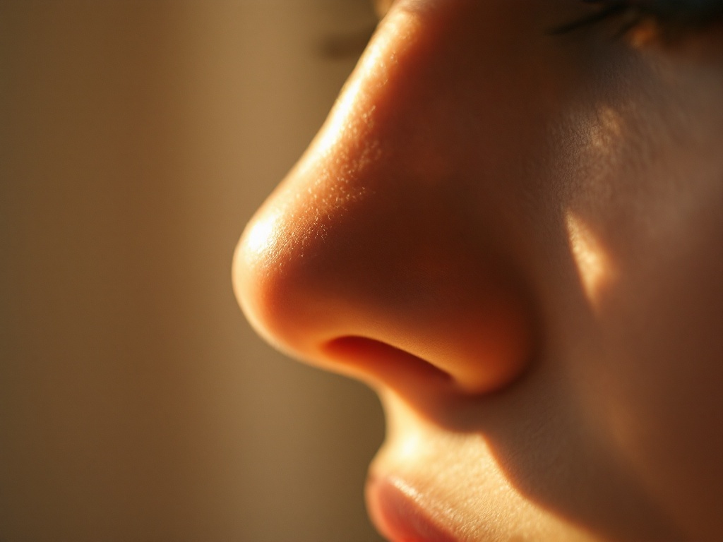
The tip is structurally different from the rest of the nose. The upper third of the nose sits over bone, and the middle third over relatively thin cartilage covered by a modest soft tissue envelope. The lower third, the tip and the area just above it known as the supratip, is covered by skin that is thicker, more sebaceous, and more richly supplied with fibrofatty tissue. Thicker skin holds more fluid, plain and simple. Postoperative edema, which is essentially protein-rich fluid trapped in the tissue, has more room to accumulate in the tip than anywhere else on the nose.
Lymphatic drainage is the second half of the story. The lymphatic channels of the nose run downward and laterally, draining toward the cheeks and eventually the lymph nodes of the neck. Surgical dissection, whether open or closed, interrupts some of these channels. In open rhinoplasty, the columellar incision and the elevation of the skin envelope transect small lymphatics at the very point where fluid needs to exit the tip. The body rebuilds these channels, but the process takes months, not weeks. Until drainage capacity recovers, fluid clears from the tip slowly, which is why the supratip can look full or slightly rounded, a temporary state sometimes described as a soft tissue polly beak.
Scar maturation adds a third layer. Wherever tissue planes are separated during surgery, the body lays down collagen to knit them back together. Early scar is disorganized, bulky, and stiff. Over roughly 12 to 18 months, that collagen remodels: fibers realign, excess matrix is broken down, and the tissue softens and thins. During this remodeling phase, the tip can feel firm or numb to the touch, and subtle definition, the small shadows and highlights that make a tip look sculpted, only emerges as the envelope shrinks down onto the cartilage framework beneath.
What actually affects the timeline? Several variables are well documented in the surgical literature. Skin thickness is the biggest one. Patients with thin skin may see near final tip definition by 6 to 9 months, while patients with thick, sebaceous skin commonly need 18 to 24 months, and occasionally longer. Revision surgery slows things further, because scarred tissue has already compromised lymphatics and less elastic skin. The extent of tip work matters too: significant cartilage reshaping, grafting, and wide dissection produce more edema than conservative refinement. Open approaches are often said to swell longer than closed approaches, and while the difference is real in the early months, most comparative data suggest outcomes converge by the one year mark.
What helps, and what is oversold. Head elevation in the first weeks, avoiding strenuous exercise early, limiting sodium, and not sleeping face down are all mechanically sensible: they reduce hydrostatic pressure and fluid delivery to the healing tissue. Nighttime taping of the supratip is used by many surgeons in thick skinned patients to apply gentle external compression while the envelope contracts, and while high quality trial data are limited, the mechanism is plausible and the intervention is low risk. Some surgeons use small, dilute steroid injections into the supratip when fullness persists beyond a few months, which works by suppressing inflammation and reducing collagen deposition. This is a legitimate tool but a double edged one: overuse can thin the skin or create depressions, so it belongs in the hands of the operating surgeon, not on a patient wish list. Lymphatic massage is frequently promoted online. Gentle techniques are unlikely to harm, but aggressive manipulation of a healing tip can theoretically shift grafts or irritate tissue, so any massage protocol should come from the treating surgeon.
When persistent fullness is not just swelling. True swelling improves, even if slowly, and it often fluctuates: worse in the morning, worse after salt or alcohol, better by evening. Fullness that is rock hard, completely static after a year, or accompanied by a visible step above the tip may instead reflect excess scar tissue or a structural issue such as inadequate cartilage support, and those are conversations to have with the surgeon rather than problems that time alone will solve.
The practical takeaway is one of calibrated patience. The tip is the thickest skinned, most heavily dissected, and most drainage dependent part of the nose, so it is mathematically last in line to reveal the final result. Judging a rhinoplasty at three months is like judging a marathon at mile ten. Photographs taken at consistent angles and lighting every few months tell the real story, and in the large majority of cases, that story is steady, quiet improvement long after the patient has stopped expecting it.
Related reading: Why the Nasal Tip Is the Last Thing to Settle After Rhinoplasty and Why the Nasal Tip Is the Last Thing to Look Right After Rhinoplasty.