
Before You Decide · February 9, 2026 · 6 min · By Gideon Maravilla
Rhinoplasty Computer Imaging: Expectations vs. Reality
How digital imaging shapes surgeon-patient dialogue before surgery.
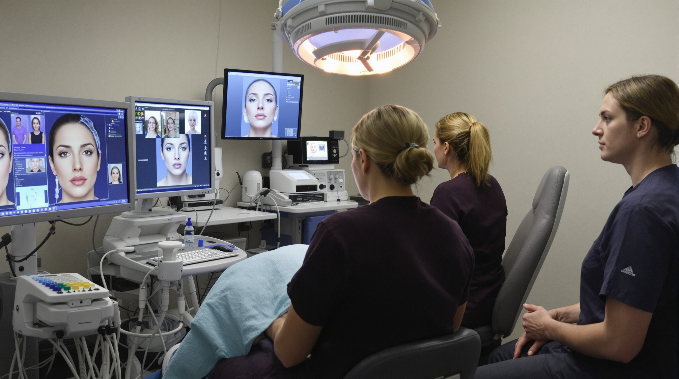
Rhinoplasty computer imaging has become standard in consultation rooms across the country, offering patients a visual framework for discussing surgical goals. The technology allows surgeons and patients to explore potential changes to dorsal height, tip projection, and profile angles in real time, creating a shared language around abstract anatomical concepts. Yet the role of imaging in rhinoplasty remains misunderstood by many candidates, who sometimes conflate the rendered image with a surgical guarantee.
Modern imaging systems use either 2D manipulation software or 3D volumetric reconstruction, each with distinct clinical applications. 2D systems allow quick exploration of profile and frontal changes by altering lines and contours on existing photographs. A surgeon might extend the tip projection by five millimeters or reduce dorsal hump height to demonstrate immediate visual feedback. These tools are intuitive and help patients grasp relative proportions without requiring extensive explanation. 3D systems, by contrast, reconstruct the nose from multiple angles and can simulate how changes in one dimension affect others, providing a more anatomically complete preview.
The critical limitation of both approaches is that they represent soft tissue behavior as static. In reality, cartilage, bone, and overlying skin respond to surgical manipulation in ways that cannot be perfectly predicted before the operation. Thick skin may not drape as smoothly as the algorithm assumes. Cartilage may spring back unpredictably after reduction. Scar tissue formation, individual healing variability, and the complex biomechanics of nasal support all introduce variance between the rendering and the final result. Surgeons who rely too heavily on imaging to set patient expectations often encounter disappointment in the months following surgery.
When used appropriately, imaging serves a narrower but valuable purpose: identifying patient concerns and communicating surgical strategy. If a patient points to a bulbous tip on the screen and says, "I want this refined," the surgeon understands the target and can explain whether that specific change is achievable given the patient's anatomy and skin type. Realistic expectations and imaging form the foundation of this discussion, and responsible surgeons frame renderings as directional rather than contractual. Some practices now explicitly document that imaging is illustrative, not predictive, as a protective and clarifying measure.
Research comparing imaged predictions to actual surgical outcomes reveals accuracy rates between 60 and 80 percent depending on the specific parameter measured and the software used. Dorsal height reduction tends to track reasonably well; tip rotation and refinement show greater variance. This gap reflects the inherent unknowns in predicting how individual tissue behaves. Patients who enter surgery understanding this variability tend to have better satisfaction outcomes than those who treat the image as a blueprint.
The imaging consultation also serves as a reality check for unrealistic requests. A patient may ask for changes that would compromise nasal function or create an appearance incongruous with facial proportions. Running those changes through imaging often helps the patient see the mismatch and recalibrate their goals. Conversely, when imaging demonstrates that a modest change produces meaningful improvement, it can help cautious patients commit to surgery they are considering. The tool amplifies communication in both directions.
For surgeons, imaging can organize pre operative planning. Measuring specific angles or distances on the rendered result provides numeric targets for intraoperative decisions. Some surgeons use imaging data to guide suture placement, cartilage reduction, and grafting strategies. Others find the numbers less useful than the visual sense of balance and proportion the imaging creates. Either way, the image becomes a reference point during the operation, particularly if the surgical plan involves multiple coordinated changes.
The emergence of AI-assisted imaging is adding another layer to this landscape. Machine learning models trained on large datasets of before and after photos can now generate predictions that incorporate population level patterns of healing and tissue response. Early adoption suggests these models may outperform manual imaging in certain scenarios, though the technology remains too new for broad clinical validation. Before and after realistic examples remain the most powerful visual teaching tools, as they represent actual healing outcomes rather than algorithmic projections.
Clinicians and patients both benefit from understanding imaging as a communication tool rather than a prediction engine. It clarifies goals, identifies anatomical constraints, and creates a visual record of the pre operative discussion. It does not, and should not, be presented as a guarantee of the final result. The best imaging consultations end with explicit acknowledgment that the nose will change during healing, that some aspects may differ from the rendering, and that the surgeon will make real time judgments during surgery based on tissue quality and unexpected findings. That framing sets the stage for realistic satisfaction with outcomes.
More in Field Notes
View all →- The nasal cycle: why the blocked side switches, and the two week log that settles it
- A Three Part At Home Test for Where Your Nose Is Actually Blocked
- Nasal Taping After Rhinoplasty: What the Ritual Does and Does Not Do
- Alar Retraction: Why Nostril Rims Pull Up After Rhinoplasty and How It Is Corrected